Point
5
Intro
 You
should recall from the spinal cord module that
proprioceptive information from muscle spindles (Ia,
II) and Golgi tendon organs (Ib) reaches the
cerebellum via the dorsal spinocerebellar
tract. The cells of origin of this tract lie in the
ipsilateral Clarke's column. This column of cells is present
only at spinal cord levels C8L3. Central processes of
dorsal root neurons that enter caudal to L3 have to ascend
to reach L3. Consequently, Clarke's column is quite enlarged
caudally. (Clarke's column neurons at L3 need to serve not
only entering fibers at L3, but all of those entering below
L3.)
You
should recall from the spinal cord module that
proprioceptive information from muscle spindles (Ia,
II) and Golgi tendon organs (Ib) reaches the
cerebellum via the dorsal spinocerebellar
tract. The cells of origin of this tract lie in the
ipsilateral Clarke's column. This column of cells is present
only at spinal cord levels C8L3. Central processes of
dorsal root neurons that enter caudal to L3 have to ascend
to reach L3. Consequently, Clarke's column is quite enlarged
caudally. (Clarke's column neurons at L3 need to serve not
only entering fibers at L3, but all of those entering below
L3.)
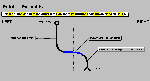 Ia, Ib and type II axons of the dorsal root
ganglia rostral to Clarke's column (C8) pass
rostrally to reach the ipsilateral caudal medulla,
where they end within the ACCESSORY CUNEATE
("wedgeshaped") NUCLEUS. This nucleus, which is
somewhat difficult to see, lies dorsal to the spinal tract
and nucleus V and lateral to the most rostral pole of
nucleus cuneatus. Cells in the accessory cuneate nucleus
send their axons to the IPSILATERAL CEREBELLUM via a
fiber bundle called the inferior cerebellar peduncle
(together with the dorsal spinocerebellar fibers). This
pathway is called the CUNEOCEREBELLAR TRACT.
Ia, Ib and type II axons of the dorsal root
ganglia rostral to Clarke's column (C8) pass
rostrally to reach the ipsilateral caudal medulla,
where they end within the ACCESSORY CUNEATE
("wedgeshaped") NUCLEUS. This nucleus, which is
somewhat difficult to see, lies dorsal to the spinal tract
and nucleus V and lateral to the most rostral pole of
nucleus cuneatus. Cells in the accessory cuneate nucleus
send their axons to the IPSILATERAL CEREBELLUM via a
fiber bundle called the inferior cerebellar peduncle
(together with the dorsal spinocerebellar fibers). This
pathway is called the CUNEOCEREBELLAR TRACT.
The accessory cuneate nucleus is concerned with relaying proprioceptive information from the arm (and neck) to the cerebellum, and the nucleus can be considered as the rostral equivalent of Clarke's column.
![]() REMEMBER: Accessory cuneate
nucleus:
REMEMBER: Accessory cuneate
nucleus:
- lies in the medulla.
- receives UNCROSSED fibers from dorsal root ganglia above C8.
- receives the same kind of information that Clarke's column does.
- projects to the IPSILATERAL cerebellum via inferior cerebellar peduncle.
- is concerned with the arm, while Clarke's column is concerned with the forearm, trunk and lower extremity.