The oral cavity is formed by a bewildering array of tissues which function in or are associated with the processes that are performed with what we typically refer to as our mouth. Lecture and lab focus on the one organ found within the oral cavity, the tongue, and the glands which empty their secretory products into the oral cavity, the salivary glands. In the lab you will also have the opportunity the examine one other specialized epithelial area, the lip. The oesophagus is the first part of the alimentary canal. Its organisation is also typical for all parts of the gastrointestinal tract (GIT).
The oral cavity is divided in a vestibule, the area "outside" the teeth, and an oral cavity proper. The entire oral cavity is lined by a stratified squamous epithelium. The epithelial lining is divided into two broad types:
- Masticatory epithelium covers the surfaces involved in the processing of food (tongue, gingivae and hard palate). The epithelium is keratinized to different degrees depending on the extent of physical forces exerted on it.
- Lining epithelium, i.e. non-keratinized stratified squamous epithelium, covers the remaining surfaces of the oral cavity.
The dorsal surface of the tongue is divided by the sulcus terminalis into an oral part, the anterior two-thirds, and a pharyngeal part, the posterior one-third. The dorsal surface of the oral part has a characteristic appearance due to the presence of a large number of small projections, the lingual papillae. The epithelium of the pharyngeal part forms a somewhat irregular surface which covers the lingual tonsils.
The lingual papillae consist of a connective tissue core covered with a stratified squamous epithelium. On the basis of their appearance four types of papillae can be distinguished - filiform, fungiform, circumvallate and foliate papillae.
 Filiform
papillae
Filiform
papillae-
are the smallest and most numerous papillae. By providing the tongue with a rough surface they aid in the manipulation and processing of foods.
Prof. Oxnard brought another function to my attention, i.e. the cleaning of the surfaces of the mouth, in particular the teeth.
 Fungiform
papillae
Fungiform
papillae-
occur singly and are fairly evenly spaced between the filiform papillae. Their connective tissue core is richly vascularised. The epithelium is slightly thinner than on the remaining surface of the tongue.
 Circumvallate
papillae
Circumvallate
papillae-
are the largest and least numerous papillae - in humans there are between 8 and 12 of them. They occur in depressions of the surface of the tongue and are surrounded with a trench formed by the infolding of the epithelium. Taste buds are particularly numerous on the lateral surfaces of these papillae. The excretory ducts of serous glands open into the trenches surrounding the papillae ("rinsing glands" or glands of von Ebner).
- Foliate papilla
-
are not well developed in humans and may be absent in aged individuals. If present, they form lamellae along the posterior and lateral border of the tongue.
The epithelium of the dorsal surface of the tongue rests on a fairly dense layer of connective tissue, which connects the epithelium firmly with the underlying muscular and connective tissues.
The muscles of the tongue (skeletal muscle) are organized into strands oriented more or less perpendicular to each other. Their actions provide the tongue with the necessary motility to participate in the formation of speech and to aid in the initial processing of foods. Control of the movement of the tongue muscles and the collection of sensory information necessitate a profuse innervation of the tongue in which a number of the cranial nerves participate (trigeminal V - sensory - anterior two-thirds, facial VII - taste, glossopharyngeal IX - sensory/taste - posterior one-third, hypoglossal XII - motor).
 Lab:
Slide TONGUE HUMAN H&E. This section illustrates
the general organisation of the tongue. Taste buds will be rare, if present
at all, in this section. Examine the tissue and have a close look at the small
salivary glands located in the connective tissue of the tongue. You should
be able to identify mucous acini and serous demilunes (see
below) attached to the acini.
Lab:
Slide TONGUE HUMAN H&E. This section illustrates
the general organisation of the tongue. Taste buds will be rare, if present
at all, in this section. Examine the tissue and have a close look at the small
salivary glands located in the connective tissue of the tongue. You should
be able to identify mucous acini and serous demilunes (see
below) attached to the acini.
Draw a part of the section in which papillae on the surface of the tongue, muscular tissue and possible some of the glands embedded between the muscular tissue of the tongue are visible.
Taste Buds
Taste buds are most numerous in the fungiform, circumvallate and foliate papillae. In addition, taste buds are found in the palate, palatoglossal and palatopharyngeal arches and in the pharyngs and larynx.
In histological sections they appear as ovoid lightly stained bodies, which extend perpendicular from the basement membrane to a little opening formed in the epithelium, the taste pore. The elongated cells that form the taste bud can functionally be divided into three groups: sensory cells, supporting (or sustentacular) cells, and basal cells. Sensory cells extend microvilli into the taste pore. These microvilli contain the receptors for the different basic taste modalities (sweet, salty, bitter and acid). Basal cells regenerate the two other cell
types.
Cell turnover is quite high, and it is thought that the cells of the taste buds are replaced (on average) every 10th day.
Lab: Slide TASTE BUDS Al. BLUE/VG SHEEP or VALLATE PAP. H&E SHEEP. Find and inspect the taste buds embedded in the epithelium of the lateral walls of the circumvallate papillae. The taste pore may not always be visible (outside the plane of section). Now look at the bottom of the trench surrounding the circumvallate papillae. Sometimes it is possible to find a duct opening into the trench. If the actual opening is outside the plane of section it is usually possible to find a section of the duct in the underlying connective tissue. Slightly deeper in the
connective tissue you may be able to identify the serous glands, which rinse the trenches surrounding the circumvallate papillae.
Draw a part of the tissue in which these structures (as many as possible) are visible.


Saliva is a mixed secretion, which is derived from numerous large and small salivary glands that all open into the oral cavity. Small salivary glands are situated in the connective tissue beneath the epithelia lining the oral cavity, and, in the case of the tongue, they may also be found between the muscular tissue. Depending on the localisation they are grouped into lingual, labial, buccal, molar and palatine glands.
The large salivary glands form three paired groups:
- the sublingual glands, which are positioned beneath the tongue and embedded deeply in the connective tissue of the oral cavity,
- the submandibular glands and
- the parotid glands, which lie outside the oral cavity.
All of these glands are tubuloacinar glands, i.e. they have secretory acini but the first part of the duct system originating from the acini also participates in the secretory process. The salivary glands are divided by connective tissue septae into lobes, which are further subdivided into lobules.
Functionally the secretory acini can be divided into two groups: those that secrete a rather liquid product - serous acini, and those that secrete a very viscous product - mucous acini. This functional differentiation is reflected in the appearance of these acini in histological sections.
- The cells forming the serous acini contain a round or slightly ovoid nucleus which is placed basally in the cell. In an H&E stain, the apical cytoplasm may appear pinkish/red or, in well-preserved tissue, contain reddish granules. The granules represent the vesicles which contain the secretory products of the cell. The digestive enzyme α-amylase is also secreted by the acinar cells.
- The cells forming the mucous acini usually contain flattened nuclei which appear to be "pressed" against the basal surface of the cell. Secretory vesicles fill much of the apical cytoplasm. The secretory product has either been dissolved during the staining process or remains unstained. Small amounts of cytoplasm which remain between the vesicles gives the apical part of the cell a distinct "spongy" appearance.
 Occasionally,
and in particular in glands located relatively close to the oral cavity, serous
cells and mucous cells may form compound or mixed acini. The
serous cells form in these cases small half-moon
or crescent-shaped structures, which attach to mucus producing acini
and empty their secretory product into interstices between the mucus-producing
cell. Following their appearance they are called serous
demilunes.
Occasionally,
and in particular in glands located relatively close to the oral cavity, serous
cells and mucous cells may form compound or mixed acini. The
serous cells form in these cases small half-moon
or crescent-shaped structures, which attach to mucus producing acini
and empty their secretory product into interstices between the mucus-producing
cell. Following their appearance they are called serous
demilunes.
Both serous and mucous acini and parts of the secretory duct system are surrounded by myoepithelial cells which by their contraction participate in the secretory process. They are usually difficult to distinguish in histological sections.
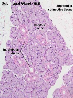
Glands located close to the oral cavity have mainly mucous secretions, whereas glands located further away from the oral cavity have mainly serous secretions. Following this general rule, the parotid glands contain almost exclusively serous acini, the submandibular glands contain both serous and mucous acini, and the sublingual glands contain mainly mucous acini or mucous acini with serous demilunes.
Ducts of the Salivary Glands
The ducts of the salivary glands can, according to their position in relation to the lobes and lobules of the glands, be divided into two parts. Interlobular and interlobar ducts are embedded in the connective tissue surrounding the lobes and lobules of the glands. Intralobular ducts are located in between the secretory acini within the lobules and, consequently, only surrounded by scant, if any visible connective tissue.
Interlobar and interlobular ducts function mainly in the conduit of the saliva and are formed by a stratified cuboidal or stratified columnar epithelium. The epithelium is replaced by the stratified squamous epithelium as they approach the opening into the oral cavity.
The product of serous glands is extensively modified by the initial part of the duct system. Intralobular ducts can on the basis of their function be divided into intercalated ducts and striated ducts. The secretory acini empty into intercalated ducts which merge into the striated ducts.
- Cells forming the intercalated ducts add bicarbonate ions to the saliva (buffering function) and absorb chloride from the saliva. They are typically formed by cuboidal epithelium.
- Striated ducts are formed by columnar cells. In contrast to many other columnar epithelia, the nucleus of these cells is located approximately midways between the apical and basal cell surfaces. The striations of the striated duct are found in the basal part of the cytoplasm of the cells where numerous mitochondria are found between infoldings of the basal cell membrane. This specialisation provides the cell with the necessary energy and surface area to perform its task in the modification of the saliva - the secretion of potassium and the absorption of sodium. Cells of the striated ducts also take up a secretable form of antibodies and release them into the
saliva.
 Intercalated ducts are difficult to identify in mucous glands and striated ducts are absent in purely mucous glands. Following the main secretory product of the major salivary glands, well-differentiated intercalated and striated ducts are a prominent feature of the parotid glands, rare in the submandibular glands and absent in the sublingual gland. An additional feature that may aid in the identification of the parotid gland are fairly large amounts of adipose tissue which is found between the secretory tissue of the lobules.
 Lab:
Slides PAROTID GLAND H&E, SUBMANDIBULAR
GLAND H&E and SUBLINGUAL GLAND H&E.
Inspect these slides. Try to identify serous and mucous acini, intercalated
ducts and, if present, striated ducts within the lobules, and excretory ducts
in the connective tissue between the lobules.
Lab:
Slides PAROTID GLAND H&E, SUBMANDIBULAR
GLAND H&E and SUBLINGUAL GLAND H&E.
Inspect these slides. Try to identify serous and mucous acini, intercalated
ducts and, if present, striated ducts within the lobules, and excretory ducts
in the connective tissue between the lobules.
High magnification images of secretory acini and ducts
of the parotid and sublingual gland are available on the MB
140 - Histology - Epithelia and Glands page. The slides of one or maybe
two of the glands do not completely conform to textbook descriptions. Which
ones, and why?.
Draw part of the tissues of the glands in which the structures
characteristic for each major salivary gland are illustrated.


Throughout the remainder of the digestive system, the histological composition of the alimentary canal can be described by the following blue-print:
- The lumen is lined by an epithelium, which rests on a vascular connective tissue, the lamina propria. The lamina propria is in turn surrounded by a narrow band of smooth muscle (muscularis mucosae). These three tissues are collectively referred to as the mucosa of the alimentary canal.
- Beneath the mucosa we find a wider zone of loose connective tissue, the submucosa, which in addition to vessels contains a nerve plexus (submucosal plexus or Meissner's plexus), which is one of the two plexi innervating the alimentary canal.
- The submucosa is surrounded by smooth muscle, which is typically divided into two differently oriented layers - an inner circular and an outer longitudinal layer. These layers of muscle are together referred to as the muscularis externa. Between two muscle layers we find the second nerve plexus innervating the alimentary canal (myenteric plexus or Auerbach's plexus).
- The alimentary canal is delimited from other tissues by a layer of loose connective tissue, the adventitia. In the case of the intraperitoneal parts of the alimentary canal, i.e. those parts which are suspended in the peritoneal cavity, a simple squamous epithelium, the serosa, delimits the adventitia from the peritoneal cavity.
Glands may be present in some parts of the wall of the alimentary canal canal. These glands are called mucosal glands if they are located luminal (or superficial) to the muscularis mucosae. If the glands extend into the submucosa they are called submucosal glands.
In the oesophagus the mucosa is formed by a stratified squamous epithelium (non-keratinised) and well defined lamina propria and muscularis mucosae. The muscularis externa is somewhat unusual in that it contains striated muscle in its upper one third, a mixture of striated muscle and smooth muscle in its middle one-third and smooth muscle in its lower one-third. Only the lowest part of the oesophagus (approx. the lowest 2 cm) enters the peritoneal cavity. With the exception of this lowest part where a serosa forms the outermost part of the adventitia, the adventitia consists
only of a layer of loose connective tissue.
Oesophageal glands are located in the submucosa. These submucosal glands produce a mucous secretion, which lubricates the epithelium and aids the passage of food. In the part of the oesophagus closest to the stomach there may be mucosal mucus-producing glands, which resemble those in the adjacent mucosa of the stomach.
They are called cardiac glands in the stomach and this name is also used for mucosal glands in the adjacent part of the oesophagus.
 Lab: Slide TRACHEA HUMAN H&E and OESOPHAGUS HUMAN H&E. Identify the different tissues that form the wall of the alimentary canal. The two slides differ in the types of muscle present in the muscularis externa and in the organisation of the muscularis mucosae. It may be difficult to identify parts of the nerve plexus innervating the oesophagus, and oesophageal glands are not present in all sections. Look for these structures, but don't get too upset if you do not
find them.
Lab: Slide TRACHEA HUMAN H&E and OESOPHAGUS HUMAN H&E. Identify the different tissues that form the wall of the alimentary canal. The two slides differ in the types of muscle present in the muscularis externa and in the organisation of the muscularis mucosae. It may be difficult to identify parts of the nerve plexus innervating the oesophagus, and oesophageal glands are not present in all sections. Look for these structures, but don't get too upset if you do not
find them.
Draw a section of the wall of the esophagus. Identify in your drawing the main parts of the wall of the alimentary canal (mucosa, submucosa, muscularis externa and adventitia).
When we think of lips we usually only think of a small part, the vermilion border (or prolabium), of the "anatomical" lips, which comprise the entire fleshy fold surrounding the oral orrifice. The outside and inside of the lips are lined by skin and oral mucosa respectively. Between the two, we find labial vessels, nerves, the orbicularis oris muscle (striated), which shapes the lips, and labial salivary glands.
The vermilion border is the area of transition from the skin to the oral mucosa. The epithelium is somewhat thicker than in other parts of the facial skin. Connective tissue papilla extend deep into the epithelium and are heavily vascularized. It is the proximity of these vessels to the surface of the epithelium which gives the prolabium it's red appearance.
 Lab:
Slide LIP HUMAN H&E. Hairs and glands (aside
from a few sebaceous glands) are usually not present beneath the epithelium
of the prolabium. Find the point of transition to the stratified squamous
epithelium of the oral mucosa. In the connective tissue beneath the oral mucosa
you may be able to find the small labial salivary glands.
Lab:
Slide LIP HUMAN H&E. Hairs and glands (aside
from a few sebaceous glands) are usually not present beneath the epithelium
of the prolabium. Find the point of transition to the stratified squamous
epithelium of the oral mucosa. In the connective tissue beneath the oral mucosa
you may be able to find the small labial salivary glands.
If you can not find them in the section, try to find them in your mouth by palpating the oral surface of the lips with the tips of your tongue - the glands are the small nodules that you feel. The sebaceous glands present in the section are very pretty - have a look - it should become clear(er) why they are alveolar glands. The thin skin present in the section is also quite stunning. The spines of the spiny cells in the stratum spinosum are actually very clearly visible.
Draw a survey of the lip and identify the prolabial and oral sides, the orbicularis oris muscle and, if present, glands embedded in the connective tissue beneath the two sides of the lip.