more about .....
Bone Dynamics
by Lis Mosekilde, M.D., D.Med.Sc., Institute of Anatomy, University of Aarhus, Denmark
Introduction
In this text, a brief description of the dynamics of bone tissue will be given - forming a basis for understanding the mechanostat theory of the human skeleton. With this knowledge, prevention/management of the "silent" bone disease - osteoporosis - should be improved.
Bone Biology
The dynamics of bone involve three different processes:
- Growth
- Modelling
- Remodelling
During childhood and the early years of adulthood, while the epiphyses are still open, the skeleton grows in length (growth), and the bones expand in diameter and achieve their external shape (modelling). During bone modelling, osteoblasts and osteoclasts work independently of each other and on different bone surfaces - often over large surface areas. The net balance is positive (i.e. there is increased bone mass) and bones reach their final external form and high bone density during this period. Both the growth and the modelling processes are controlled by hormones and by mechanical forces - mechanical usage.
Around the age 20-25 years, peak bone mass is achieved as a result of these processes. Individual peak bone mass is dependent on genetic, racial and hormonal factors and also on external factors: physical activity and nutrition. Establishing a high peak bone mass gives the optimum "bone bank" from which to draw during aging. Peak bone mass is normally 20-25% higher in men than in women.
From the age of 20-25 years, bone mass begins to decline. This early age-related loss is especially detectable at places where trabecular bone dominates e.g. the vertebral bodies. Loss of bone mass with age is unavoidable and is caused by the third process - bone remodelling.
During the remodelling process, osteoclasts and osteoblasts work closely together in time and space (coupling), and they work in units called BMU (Bone Multicellular Units). When a remodelling process has just been completed, a small amount of bone (trabecular or cortical) has been removed and has been replaced by new bone. The process seems designed to renew old bone - possibly having microfractures or with dead osteocytes - and to reorganize trabecular structures to achieve the maximum strength obtainable with the bone mass remaining available. The remodelling process has another role: it ensures the maintenance of calcium homeostasis. Trabecular bone is remodelled much more rapidly than cortical bone (5-10 times more rapidly). The remodelling process is itself governed by hormonal and mechanical stimuli - as is also seen in the growth and modelling processes.

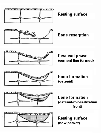
Figure 1:
Drawing showing the different phases in the bone remodelling
process/cycle.
Each remodelling process is initiated by an Activation (A) by which osteoblastic lineage cells
start to secrete collagenase which removes the thin layer of
unmineralised bone typical of a resting bone surface. This exposes
the mineralised bone underneath to the multinucleated mobile,
osteoclasts. During osteoclastic bone
Resorption (R), Howship's lacunae are excavated to a maximum
depth of 50-60 µm. A short reversal phase, where the cement
line is formed, follows, and then bone
Formation (F) normally begins. If coupling has taken place,
osteoblasts produce osteoid (collagen and ground substance) at a
rate of 0.5-1.0 µm per day. When the osteoid thickness has
reached approximately 12-15 µm, mineralisation begins from
the bottom (mineralisation front). At the termination of each
remodelling process, the bone surface is again covered by an
extremely thin layer of non-mineralised bone and a layer of flat
lining cells. The bone is again converted into a resting surface
(Fig.
1).
| The remodelling process, which is designed to
renew old bone of inferior quality and to replace bone with
microfractures, has its own cost - the balance in adults is
normally negative (i.e. after each remodelling process there is
therefore a reduced mass). There is thus an unavoidable loss of
bone mass with age. It also has another cost: it causes disruption
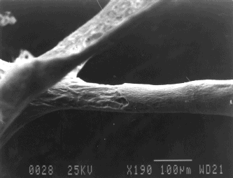
of the trabecular network with age. Because the balance is
negative, there is a thinning of the trabecular structures in the
network (Fig.
2), and this makes fortuitous osteoclastic
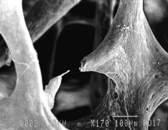
perforations possible. As the normal resorption depth is
approximately 40-50 µm, one resorption site covering more
than half of the circumference of a trabecula, or two resorption
sites, one on each side of a trabecula, could easily perforate a
trabecular structure with a diameter of 90-100 µm [90-120
µm is the normal thickness of horizontal trabeculae in the
vertebral body of elderly individuals (Fig. 3)].
The bone loss caused by the negative balance is, in theory, reversible, while bone loss caused by perforations is irreversible. |
|
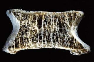
In summary, the three processes - growth, modelling, and remodelling - although different in working methods - are all governed by the same cells and are all dependent on intrinsic factors and also on mechanical stimuli. This means that the form, size, density, and strength of the skeleton at any age are dependent on its mechanical usage (Figs. 4 & 5).
 |
 |
Figure 4: A vertebral body from a young individual. |
Figure 5: A vertebral body from an elderly person |
The Mechanostat
The skeleton acts as a mechanostat: bone mass and strength will throughout life be regulated to meet the mechanical demands. Lack of physical activity (disuse) leads to loss of bone mass, mainly through the remodelling process. On the other hand, vigorous exercise (overuse) will increase bone mass, size and strength (mainly through the modelling process by which new bone is added on external bone surfaces).
The osteocytes have a key role and function for the mechanostat: every movement, lift, step, jump will be registered by the osteocytes lying in their tiny lacunae within the bone tissue. They will sense the stress or strain on bone and send the signals to the active bone cells on the surface (osteoblasts and osteoclasts). The pathway of these signals has been extensively studied in cell culture, in animal studies and in humans.
Furthermore, clinical studies in humans have shown that:
- Exercising young people have higher bone mass than their sedentary counterparts.
- The dominant arm of tennis players is much larger and stronger than the non-dominant arm.
- Exercise programmes can increase bone mass at all ages.
- Strict bed rest leads to loss of 1-2% trabecular bone per week.
Osteoporosis
Osteoporosis is a state (disease) with decreased bone mass and microarchitectural deterioration of bone, leading to fractures without major trauma.
Osteoporosis leads to fractures of the spine, the wrist and the hips. It thereby causes acute and chronic pain and can lead to invalidation.
Osteoporosis is the most common metabolic bone disease in the industrialised world. It behaves like an epidemic: it has spread from Europe and USA to Asia and the Middle East. It used to be a women's disease, but it is now just as much a disease of men.
Osteoporosis is often treated as though it were a hormonal disease alone (lack of estrogen after the menopause), but the epidemic character and the fact that men are seriously affected too shows that the main cause is totally different: lack of exercise.
As explained above, the skeleton behaves like a mechanostat - and as the physical demands decrease with adaptation to the modern lifestyle, so the mechanostat will react, and bone mass and strength will decrease to a very low level.
Summary
Bones are living structures which undergo dynamic changes throughout life. The four different bone cells each play a role concerning bone turnover. The balance in this turnover is determined by hormonal factors and mechanical usage. However, it is only recently that the importance of mechanical usage and the mechanostat theory have been acknowledged and accepted. Hopefully, this will lead to new effective preventive strategies against osteoporosis - one of which will almost certainly be exercise.

