Lymphoid (or lymphatic) tissues, which mainly consist of dense accumulations of lymphocytes, are widely distributed in the body. Lymphoid tissues are typically located at sites that provide a possible route of entry of pathogens and/or sites that are liable to infections. Epithelia delimit all other tissues from the "outside world" and it is not surprising that lymphoid tissues are often found near them. Such lymphoid tissues are grouped together as epithelium-associated lymphoid tissues. Depending on their precise location these lymphoid tissues may be referred to as e.g. mucosa-associated lymphoid tissue (MALT) or bronchus-associated lymphoid tissue (BALT). The tonsils or Peyer's patches are examples of mucosa-associated lymphoid tissues. Lymphoid tissues represent the sites of proliferation and differentiation of lymphocytes.
Lymphoid organs may be defined as anatomical "entities" which consists chiefly of lymphoid tissues. The thymus is a primary lymphoid organ in that it supplies other lymphoid organs and tissues with T-lymphocytes. Inserted into the blood and lymph vascular system, the spleen and lymph nodes (secondary lymphoid organs) monitor the internal environment of the body.
The Thymus (Greek: thymos - sweetbread) is situated in the upper parts of the thorax, behind the sternum and the upper four costal cartilages, in the anterior and superior mediastina. The size of the thymus changes in the course of life. It weighs about 10-15 g at birth and reaches its top weight (about 30-40 g) at puberty. After puberty a progressive involution (see below) occurs, which leaves a middle-aged person with a thymus weighing about 10 g. The thymus consists of a right and left lobe which are joined by connective tissue.
The thymus is enclosed by a thin connective tissue capsule from which numerous septae extend into the thymus subdividing the two lobes into numerous lobules (about 0.5 -2 mm in diameter). Blood vessels enter and leave the thymus via the connective tissue septae. Each lobulus is divided into a darker peripheral zone, the cortex, and a lighter, central zone, the medulla. Medullary tissue is continuous from lobule to lobule throughout each lobe.
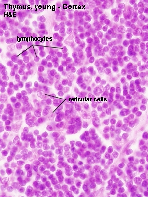
Reticular cells and macrophages are present in addition to the lymphocytes, which are the dominant cell type within the lobules.
- Reticular cells
-
are quite abundant. Their cytoplasm is eosinophilic, and their large, ovoid and light nuclei may contain one or two nucleoli. The cells are branched, and their slender processes are connected with the processes of other reticular cells to form a cellular reticulum (or cellular network). This cellular network (reticular fibres are scant in the thymus) provides support for other cells of the thymus.
Reticular cells ensheath the cortical capillaries; they form an epitheloid layer which delimits the cortical tissue from the connective tissue and secrete substances (hormones and other factors) important for thymic function. Thereby they create and maintain the microenvironment necessary for the development of T-lymphocytes in the cortex. Their functions thus go beyond those of "typical" reticular cells and, to reflect this, they are also referred to as thymic epitheliocytes.
- Macrophages
-
occur in both cortex and medulla. They are difficult to distinguish from the reticular cells in out preparations.
- Lymphocytes
-
are present in both cortex and medulla, but are more numerous (denser) in the cortex. Their sizes are variable (5 - 15 µm) in the cortex but generally small in the medulla. The vast majority of them will be developing T-lymphocytes. They are also called thymic lymphocytes or thymocytes.
 Lab: Slide THYMUS YOUNG HUMAN H&E. Identify the connective tissue capsule and septae, a lobule and its cortex and medulla at low magnification. Identify lymphocytes and thymic corpuscles. They look pretty much like a sliced (very, very small) onion. Take a close look at the medulla and try to find some cells which contain large and light nuclei. They will be either macrophages or reticular cells.
Sketch part of the thymus at low magnification. Identify medulla and cortex. Draw a segment of a lobule at high magnification and identify lymphocytes and nuclei of reticular cells/macrophages and, if possible a Hassal corpuscle.

Function of the Thymus
The thymus is necessary for the development of the recirculating pool of small, long-lived (in humans many years) lymphocytes, the T-lymphocytes, which are mainly responsible for the cell-mediated part of an immune response. Stem cells invade the cortical regions of the thymus, where they divide to form lymphocytes. Only a small fraction (estimates range from 10-30%) of the cells generated in the cortex leave the thymus. They migrate via the medulla into the blood stream to populate the T-lymphocyte areas of other lymphoid tissues and organs. Cells which react incorrectly
towards "self-antigens" die and are removed by cortical macrophages.
Since the function of the thymus is to produce lymphocytes for the other lymphoid tissues it is a primary lymphoid organ.
After puberty much of the parenchyma of the thymus, in particular cortical lymphoid tissue, is replaced by adipose tissue. The process, which is called involution, initially proceeds rapidly but slows down in adulthood. Involution is under the control of steroid hormones (both sexual hormones and stress hormones). Although most pronounced in the thymus, involution is a common feature of all lymphoid tissues.
Another age-related phenomenon is the increase in size of the thymic (or Hassall's) corpuscles. Thymic corpuscles are rounded eosinophilic structures, which consist of concentrically arranged, flattened cells. Thymic corpuscles are likely to be formed by reticular cells. Similar structures occur also in the tonsils. The size of these structures varies from 20 µm to more than 100 µm. Thymic corpuscles may calcify, and their core may "dissolve" leading to the formation of a cyst.
Lymph nodes are small, flattened, oval or bean shaped organs, which are situated in the course of the collecting lymph vessels. Their size is variable (from a few mm to more than 2 cm). The capsule and trabeculae of lymph nodes are formed by connective tissue. Afferent lymph vessels penetrate the capsule and empty into the subcapsular space. The lymph continues thereafter through cortical and medullary sinuses towards the efferent lymph vessels, which emerge from the hilus of the lymph node. The walls of the sinuses can be traversed freely by all components of the lymph, which
allows lymphocytes to enter/leave the lymphoid tissue (as part of their constant circulation) or to get in contact with antigens/antigen-presenting cells that may arrive with the lymph.
In lymph nodes we find B- and T-lymphocytes, macrophages and reticular cells.
- Reticular cells
-
(and reticular fibres) form a delicate network between the capsule and trabeculae. Only their large and light nuclei are easily visible in the microscope (the cytoplasm is only weakly eosinophilic). Lymphocytes and macrophages are housed in the network of reticular cells and reticular fibres formed by them. The processes of reticular cells and reticular fibres extend into and criss-cross within the sinuses.
- Lymphocytes
-
which are located in the outer cortex of the lymph node are likely to represent B-lymphocytes. They are organised into spherical masses - lymphoid nodules or follicles. Sites within the cortex at which B-lymphocytes have been stimulated to proliferate (by contact with an antigen) appear lighter than the surrounding tissue and allow you to identify the centres of lymphoid nodules. The lighter stained parts of the nodules are called germinal centres. Mature B-lymphocytes (plasma cells) are located in cord-like extensions of the lymphoid tissue into the medulla, the medullary cords. T-lymphocytes are located in the more diffuse tissue between the nodules and in the paracortex, i.e. the deep part of the cortex.
- Macrophages
-
are found scattered within the lymphoid tissue. They are, once again, difficult to distinguish from the reticular cells.
Blood Vessels
Blood vessels enter the lymph nodes through the hilus and travel initially in the connective tissue trabeculae that extend from the hilus into the parenchyme of the lymph node. They continue in the medullary cords towards the cortex and give off capillaries to the surrounding tissue as they do so. Postcapillary venules (or high-endothelial venules) in the deep cortex have a characteristic low cuboidal epithelium (quite unlike the squamous epithelium that we usually would expect to see). Lymphocytes, which reach the lymph node via the blood stream, may migrate through this epithelium as part of their
recirculation. Larger venules accompany the arteriolar branches as they leave the lymph nodes.


